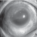
The retina is a light-sensitive sheet located at the back of the eye. We say that a retinal detachment occurs when this sheet separates from the eye wall, to which it is normally attached.
Who is most susceptible to getting it?
Retinal detachment affects approximately one in 10,000 people a year. It is a severe eye condition that can affect anyone, although it typically manifests in adults. It occurs more frequently in myopic people or in those people with family members who have suffered one. An eye bruise may also be the cause. In some rare cases, retinal detachment is an inherited disease and can occur even in children.
What are your causes?
Most retinal detachments are caused by the presence of one or more retinal tears or holes, which in turn may be due to:
- normal eye aging
- abnormal eye growth (sometimes due to near-sightedness)
- inflammation
- trauma
Normal aging can cause retinal thinning and deterioration. But more often, the cause of retinal deterioration and tears is retraction of the vitreous body, the gelatinous fluid that fills the inside of the eye. The vitreous is attached to the retina in several places on the back wall of the eye.
If the vitreous retracts, it can drag a fragment of the retina along with it and cause a tear or hole in the retina. Although such collapse of the vitreous occurs with age and does not cause any damage to the retina, it can also be caused by abnormal growth of the eye (sometimes due to myopia), inflammation, or trauma.
Once a retinal tear appears, watery fluid from the vitreous space can pass through the retinal hole and flow between the retina and the back wall of the eye. Then, the retina separates from the back wall of the eye and detaches. The portion of the retina that is detached will not function properly and a blur or black spot will appear in your vision.
What are the symptoms?
The symptoms of a retinal detachment will depend on the degree of its evolution, but some of them may be the following:
- Black floating bodies, spots, or “floaters” (myodesopsias)
- Luminous flashes
- Wavy or watery vision
- A dark shadow in some areas of vision
- Blurred central vision
- Rapid vision loss
- Complete loss of vision in one eye
Most of the time, black floaters or spots and bright flashes do not indicate any serious problem and are relatively common in middle-aged and elderly people.
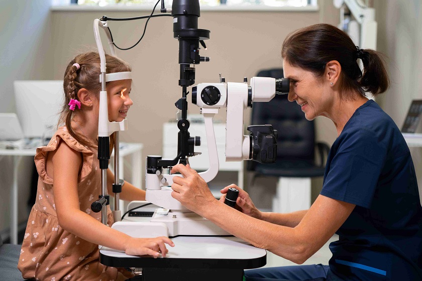
However, it is necessary for the patient to go to the emergency room so that the ophthalmologist can perform an eye examination to check if there are tears in the retina.
It is advisable by God Service Eye Clinic to perform this examination as soon as possible since recent retinal tears can be treated early with a laser before they can cause retinal detachment.
Some retinal detachments may appear without seeing floaters or flashes of light. In this case, patients may notice wavy or watery vision or a dark shadow in some areas of their vision. The subsequent development of a retinal detachment will lead to blurred central vision and significant vision loss.
Some rare retinal detachments can develop quickly. If so, the patient will experience a complete loss of vision in one eye. A rapid loss of vision may also be due to bleeding into the vitreous cavity, which may occur concomitantly if the tear ruptures a retinal vessel.
What does the treatment consist of?
If the detachment is not treated, vision is irrecoverably lost, since retinal atrophy and chronic ocular inflammation progressively occur. Over time and in certain cases, atrophy of the entire eye can even occur.
Once the retina is detached, the treatment is always surgical. The objectives of the treatment are to close the tear or break that has led to retinal detachment using laser or cryotherapy and put the retina in its place.
This can be done by pushing from outside the wall of the eyeball toward it, by placing silicone belts or pieces, or by performing a vitrectomy and then introducing gas or silicone oil into the vitreous cavity. The intervention is performed on an outpatient basis under local anesthesia and sedation.
Treat retinal detachment with a vitrectomy
Vitrectomy is a type of eye surgery for the treatment of diseases of the retina and vitreous, including retinal detachment. During a vitrectomy, the vitreous humor is removed and usually replaced with a saline solution or a bubble of gas, air, or silicone oil.
In the event that the vitreous humor is replaced by gas or silicone, it may be necessary for the patient to remain in a prone or side position for a few days.
Furthermore, if the inside of the eye is filled with gas or air, you will not be able to fly in an aeroplane or climb to a height greater than 500 meters above sea level until the bubble has disappeared since changes in altitude can affect it.
What happens during vitrectomy?
A vitrectomy is normally performed in an outpatient surgery lasting between half an hour and several hours and under local or general anesthesia to put the eye to sleep.
During surgery, the ophthalmologist makes three small cuts or incisions smaller than 1 millimeter in the white layer of the eye, the sclera, using a microscope to see the inside of the eye.
The surgeon will use small-caliber surgical instruments to perform one or more of the following steps:
- remove opacified vitreous humor
- remove scar tissue from the retina
- Clear the eye of any foreign objects.
- reposition the retina at the back of the eye
- use a laser to repair tears in the retina
- placing a gas or air bubble in the eye to help the retina stay in place (the gas bubble later disappears on its own)
- place a bubble of silicone oil in the eye (the oil is removed in a second intervention)
After the vitrectomy procedure
After the operation, the ophthalmologist will prescribe pain-relieving drugs and drops to be administered for several weeks and will tell you when you can return to your normal life.
As with any surgery, there are some risks, although they are smaller than the expected benefits to vision. Some of the risks of the procedure include hemorrhages, retinal detachment, elevated intraocular pressure, or infections.


 (3 votes, average: 3.67 out of 5)